Order Now
- Home
- About Us
-
Services
-
Assignment Writing
-
Academic Writing Services
- Urgent Assignment Help
- Writing Assignment for University
- College Assignment Help
- SPSS Assignment Help
- HND Assignment Help
- Architecture Assignment Help
- Total Assignment Help
- All Assignment Help
- My Assignment Help
- Student Assignment Help
- Instant Assignment Help
- Cheap Assignment Help
- Global Assignment Help
- Write My Assignment
- Do My Assignment
- Solve My Assignment
- Make My Assignment
- Pay for Assignment Help
-
Management
- Financial Management Assignment Help
- Business Management Assignment Help
- Management Assignment Help
- Project Management Assignment Help
- Supply Chain Management Assignment Help
- Operations Management Assignment Help
- Risk Management Assignment Help
- Strategic Management Assignment Help
- Logistics Management Assignment Help
- Global Business Strategy Assignment Help
- Consumer Behavior Assignment Help
- MBA Assignment Help
- Portfolio Management Assignment Help
- Change Management Assignment Help
- Hospitality Management Assignment Help
- Healthcare Management Assignment Help
- Investment Management Assignment Help
- Market Analysis Assignment Help
- Corporate Strategy Assignment Help
- Conflict Management Assignment Help
- Marketing Management Assignment Help
- Strategic Marketing Assignment Help
- CRM Assignment Help
- Marketing Research Assignment Help
- Human Resource Assignment Help
- Business Assignment Help
- Business Development Assignment Help
- Business Statistics Assignment Help
- Business Ethics Assignment Help
- 4p of Marketing Assignment Help
- Pricing Strategy Assignment Help
- Nursing
-
Finance
- Finance Assignment Help
- Do My Finance Assignment For Me
- Financial Accounting Assignment Help
- Behavioral Finance Assignment Help
- Finance Planning Assignment Help
- Personal Finance Assignment Help
- Financial Services Assignment Help
- Forex Assignment Help
- Financial Statement Analysis Assignment Help
- Capital Budgeting Assignment Help
- Financial Reporting Assignment Help
- International Finance Assignment Help
- Business Finance Assignment Help
- Corporate Finance Assignment Help
-
Accounting
- Accounting Assignment Help
- Managerial Accounting Assignment Help
- Taxation Accounting Assignment Help
- Perdisco Assignment Help
- Solve My Accounting Paper
- Business Accounting Assignment Help
- Cost Accounting Assignment Help
- Taxation Assignment Help
- Activity Based Accounting Assignment Help
- Tax Accounting Assignment Help
- Financial Accounting Theory Assignment Help
-
Computer Science and IT
- Robotics Assignment Help
- Operating System Assignment Help
- Data mining Assignment Help
- Computer Network Assignment Help
- Database Assignment Help
- IT Management Assignment Help
- Network Topology Assignment Help
- Data Structure Assignment Help
- Business Intelligence Assignment Help
- Data Flow Diagram Assignment Help
- UML Diagram Assignment Help
- R Studio Assignment Help
-
Law
- Law Assignment Help
- Business Law Assignment Help
- Contract Law Assignment Help
- Tort Law Assignment Help
- Social Media Law Assignment Help
- Criminal Law Assignment Help
- Employment Law Assignment Help
- Taxation Law Assignment Help
- Commercial Law Assignment Help
- Constitutional Law Assignment Help
- Corporate Governance Law Assignment Help
- Environmental Law Assignment Help
- Criminology Assignment Help
- Company Law Assignment Help
- Human Rights Law Assignment Help
- Evidence Law Assignment Help
- Administrative Law Assignment Help
- Enterprise Law Assignment Help
- Migration Law Assignment Help
- Communication Law Assignment Help
- Law and Ethics Assignment Help
- Consumer Law Assignment Help
- Science
- Biology
- Engineering
-
Humanities
- Humanities Assignment Help
- Sociology Assignment Help
- Philosophy Assignment Help
- English Assignment Help
- Geography Assignment Help
- Agroecology Assignment Help
- Psychology Assignment Help
- Social Science Assignment Help
- Public Relations Assignment Help
- Political Science Assignment Help
- Mass Communication Assignment Help
- History Assignment Help
- Cookery Assignment Help
- Auditing
- Mathematics
-
Economics
- Economics Assignment Help
- Managerial Economics Assignment Help
- Econometrics Assignment Help
- Microeconomics Assignment Help
- Business Economics Assignment Help
- Marketing Plan Assignment Help
- Demand Supply Assignment Help
- Comparative Analysis Assignment Help
- Health Economics Assignment Help
- Macroeconomics Assignment Help
- Political Economics Assignment Help
- International Economics Assignments Help
-
Academic Writing Services
-
Essay Writing
- Essay Help
- Essay Writing Help
- Essay Help Online
- Online Custom Essay Help
- Descriptive Essay Help
- Help With MBA Essays
- Essay Writing Service
- Essay Writer For Australia
- Essay Outline Help
- illustration Essay Help
- Response Essay Writing Help
- Professional Essay Writers
- Custom Essay Help
- English Essay Writing Help
- Essay Homework Help
- Literature Essay Help
- Scholarship Essay Help
- Research Essay Help
- History Essay Help
- MBA Essay Help
- Plagiarism Free Essays
- Writing Essay Papers
- Write My Essay Help
- Need Help Writing Essay
- Help Writing Scholarship Essay
- Help Writing a Narrative Essay
- Best Essay Writing Service Canada
-
Dissertation
- Biology Dissertation Help
- Academic Dissertation Help
- Nursing Dissertation Help
- Dissertation Help Online
- MATLAB Dissertation Help
- Doctoral Dissertation Help
- Geography Dissertation Help
- Architecture Dissertation Help
- Statistics Dissertation Help
- Sociology Dissertation Help
- English Dissertation Help
- Law Dissertation Help
- Dissertation Proofreading Services
- Cheap Dissertation Help
- Dissertation Writing Help
- Marketing Dissertation Help
- Programming
-
Case Study
- Write Case Study For Me
- Business Law Case Study Help
- Civil Law Case Study Help
- Marketing Case Study Help
- Nursing Case Study Help
- Case Study Writing Services
- History Case Study help
- Amazon Case Study Help
- Apple Case Study Help
- Case Study Assignment Help
- ZARA Case Study Assignment Help
- IKEA Case Study Assignment Help
- Zappos Case Study Assignment Help
- Tesla Case Study Assignment Help
- Flipkart Case Study Assignment Help
- Contract Law Case Study Assignments Help
- Business Ethics Case Study Assignment Help
- Nike SWOT Analysis Case Study Assignment Help
- Coursework
- Thesis Writing
- CDR
- Research
-
Assignment Writing
-
Resources
- Referencing Guidelines
-
Universities
-
Australia
- Asia Pacific International College Assignment Help
- Macquarie University Assignment Help
- Rhodes College Assignment Help
- APIC University Assignment Help
- Torrens University Assignment Help
- Kaplan University Assignment Help
- Holmes University Assignment Help
- Griffith University Assignment Help
- VIT University Assignment Help
- CQ University Assignment Help
-
Australia
- Experts
- Free Sample
- Testimonial
Question
Task: Prepare a report on the topic 'Fluid mosaic model of membrane structure'.
Answer
Introduction
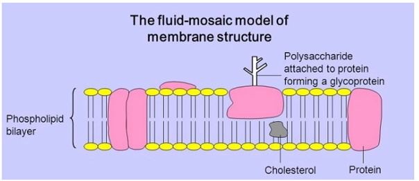
A cell membrane with the property of being a two-dimensional liquid with mixed composition is referred to as a fluid mosaic model. The hydrophobic components that are incorporated into the development of the membrane give the cell membrane its fluid quality and cause the lipids and proteins to migrate from one side of the membrane to the other. The membrane has a nature that is more fluid. The fluid mosaic model uses the term "mosaic" to describe anything that was created by combining many elements, which explains why it is appropriate for this model of biology assignment. The fact that the cell membrane is made up of several components justifies the use of the word mosaic in the model's name, the fluid mosaic model. The fluid quality of the cell membrane is caused by the presence of phospholipids, which do not form bonds with one another. The phospholipid molecules have a head that is drawn to water, causing it to point in the direction of the cell membrane's outer surface and giving it a hydrophilic quality. There are two directions that the molecules move in. The tail aids in removing the water that forms a bilayer to create non-polar hydrophobia by wading it away. Fatty acids make up the phospholipid tail, which is continually moving. After the creation of the fluid mosaic model, the development of the bilayer became known. The process is weaker since each person goes through it alone (Catala, 2012). The production of cholesterol is one of the many proteins and chemicals that are entrenched in the bilayer. The plasma membrane has a consistency similar to that of vegetable oil that has been put at room temperature, which allows proteins and several other things to travel around it. Understanding the plasma membrane is accomplished using the fluid mosaic model. The cell membrane is made up of several substances, each serving a specific function. The membrane is stabilized by the cholesterol that becomes embedded in its bilayer, preventing it from hardening at lower body temperatures. The plasma membrane of the cells found in animals is also better understood using the fluid mosaic concept. Glycoproteins and glycolipids are formed from the carbohydrate chains that make up the cell membrane's outer layer. The way that carbs are formed varies from person to person, and how they are formed relies on the type of blood that each individual has (Leabu, 2013).
How did Fluid Mosaic Model Come into Existence?
To explain the composition of the plasma membrane, S.J. Singer and G. L. Nicolson developed the fluid mosaic model. It was believed to be a fundamental component of cell membranes that would aid in future research by explaining the information already available regarding the proteins and lipids found in the membrane (Nicolson, 2016). Even though the concept has changed over time, it is still regarded as a valid theory for explaining the nano-structures of many intracellular and cellular membranes seen in the cells of both plants and animals. The arrangement of the particles in the cell membrane is depicted by the fluid mosaic model. Meanwhile, new information about lipid rafts, proteins, and glycoproteins emerged, aiding in the description of the membrane's structure. Due to the ideas mentioned to define the Fluid Mosaic Model, new information about the cell membrane came to light. The mosaic character is given more weight in recent information (Nicolson, 2016).
What are the different organelles of a cell?
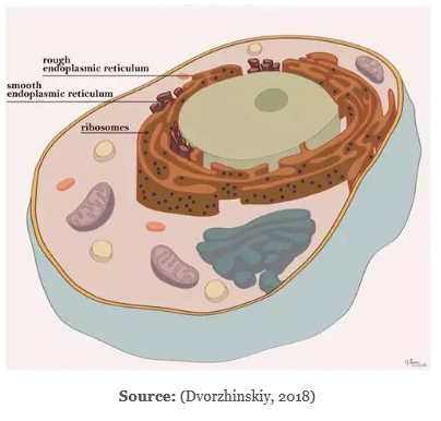
The term "cell organelles" refers to various cellular elements present in both plants and mammals. Organelles are the various structures that make up a cell. The membrane that surrounds the cell's organelles has a specific structure and function (Morange, 2013). All of a cell's parts are contained by the cell membrane, which also shields it from the environment. It also facilitates the entry and exit of ion-regulating particles from the cell. The coordination of these cell organelles is necessary for the cell to function normally. The following list of cell organelles has been discussed:
• Endoplasmic Reticulum:
This is a collection of membrane canals that is hydrated. They are viewed as a means of transportation that aids in moving materials inside the cell. A component of the endomembrane system that extends the nuclear envelope is the endoplasmic reticulum. Rough Endoplasmic Reticulum and Smooth Endoplasmic Reticulum are the two forms of the endoplasmic reticulum. Tubules, vesicles, and cisternae make up the rough endoplasmic reticulum. They aid in the synthesis of proteins and are present throughout the cell. An area of the smooth endoplasmic reticulum serves as storage. It aids in the synthesis of lipids and steroids. It also aids in cell detoxification. The Rough Endoplasmic Reticulum has several ribosomes linked to it, which results in an uneven structure and justifies the name of the organelle. The rough endoplasmic reticulum aids in the synthesis of proteins that leave the cell. These proteins are moved to the lumen inside the endoplasmic reticulum, where they undergo extensive shape modification. The Smooth Endoplasmic Reticulum receives the protein through the lumen and processes it further there. The absence of ribosomes in the Smooth Endoplasmic Reticulum contributes to its smooth structure. Due to a lack of ribosomes, it is unable to produce proteins. Making lipids and enzymes is the Smooth Endoplasmic Reticulum's primary job.
Functions of the Endoplasmic Reticulum:
• Produces and secretes steroid hormones
• Assists in the synthesis of lipids such as cholesterol and phospholipid
• Assists in the metabolism of carbohydrates
• Assists in the release of calcium ions, which is crucial for the nervous and muscular systems
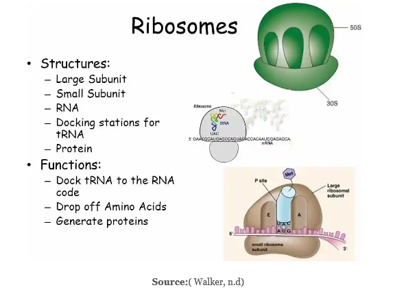
• Ribosomes: This structure is where protein synthesis occurs, producing protein and ensuring the survival of living cells. It is made up of several molecules. All cell types, including prokaryotic and eukaryotic cells, have ribosomes. Ribosomes are required by every single cell for the production of proteins. The tiny and big subunits of the ribosomes are comprised of ribosomal RNA and ribosomal proteins. The messenger RNA (mRNA) and the amino acids that are associated with the transfer RNAs (tRNAs) are transported toward the ribosome during the synthesis of proteins. Proteins are made with the aid of amino acids. The Rough Endoplasmic Reticulum is where the ribosomes are affixed. They are floating in the cytoplasm and are unbound. They have no membranes and are quite tiny in size. Two-thirds of ribonucleic acid and one-third of protein make up this substance. They are also known as prokaryotic 70s or eukaryotic 80s, depending on the type of cell they are found in. S stands for size and density. The skin, hair, eyes, and face all contain ribosomes.
Functions of Ribosomes:
• Assembles amino acids to make proteins, which are thought to be a necessary component for a cell to function. Messenger RNA (mRNA) aids in protein synthesis with the support of the nucleus and cytoplasm.
• The messenger RNA (mRNA) polymers are encircled by ribosome subunits in the cytoplasm.
• The proteins are transported outside of the cell by the newly created ribosomes.
• Golgi Apparatus: Both plant and animal cells have the Golgi complex or the Golgi apparatus in their cytoplasm. It is an organelle that resembles a flat, layered sac that changes proteins and aids in their packaging. It consists of a group of membranes that closely collaborates with the endoplasmic reticulum to change proteins and carbohydrates. It typically has 6 cisternae but can have up to 20 in total. Cells of eukaryotic organisms have the Golgi apparatus. It is surrounded by a membrane, the size of which varies between locations. It actively participates in the production, storage, and delivery of goods made by the endoplasmic reticulum. The Golgi apparatus has a pancake-like form due to the folded membranes there. It serves as the home for numerous vesicles that the Smooth Endoplasmic Reticulum produces. Before being circulated throughout the cell, the Endoplasmic Reticulum's produced proteins are processed by the Golgi apparatus. The protein enters the Golgi apparatus from one side and exits from the opposite side toward the cell's plasma membrane with the assistance of the endoplasmic reticulum. One of the recognized models of the plasma membrane is the fluid mosaic model. A cell's Golgi body might vary depending on what it does.
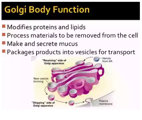
Source: ( Mandira and Kate, 2017) ( Mandira and Kate, 2017)
The Golgi complex has the following functions:
• Absorbing compounds, assisting in secretion, and forming secretory vesicles
• Assisting in the formation of enzymes
• Assisting in the production of hormones
• Assisting in the storage of proteins
• Assisting in the formation of acrosomes
• Assisting in the formation of intracellular crystals
• Assisting in the formation of milk protein droplets
• Assisting in the formation of plant cell walls
• Assisting in the secretion of glycoprotein
• Lysosomes: Enzyme sacs are what are typically referred to as lysosomes. It aids in the digestion of the various lipids and nucleic acids found in a cell. The lysosomes' internal environment has an acidic character. The circumstances found in the lysosome membrane offer the enzymes a favourable environment in which to work. The cytoplasm is where they are located. The lysosome aids in the breakdown of dying cells, organelles, poisons, and food particles. Lysosomes are frequently referred to as suicide sacks. The lysosome buds from the Golgi complex's membrane sacs. One of the functions of lysosomes is to act as a container for the removal of waste ( Pu, Guardia, Keren-Kaplan and Bonifacino, 2016). Both the prevention and the aetiology of various diseases may be affected by the chemicals that process lysosomes.
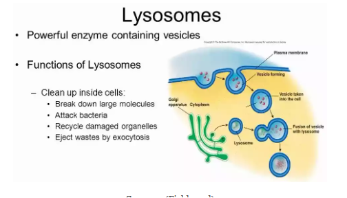
Source: (Fields, n.d)
Functions of lysosomes:
• Aid in intracellular digestion
• Remove dead cells
• Aid in metamorphosis
• Aid in protein synthesis
• Aid in fertilization
• Aid in the process of ontogenesis
• Aid in the removal of toxins
• Aid in the digestion of food particles
• Aid in the formation of bone cells
What is Kartagener Syndrome, and How is it Caused?
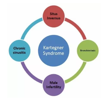
Source: (Plessis and Wahba, (n.d))
A person who has acute sinusitis, bronchiectasis, and situs inverse is said to have Kartagener syndrome. Kartagener syndrome is caused by the improper acquisition of the motor protein dynein. Chest infections and infertility result from the cilia moving improperly (Xu, Gong and Wen, 2017). Biofilms gathered from the air are incorporated into the mucus, causing bacterial infections and tissue damage. Kartagener syndrome-affected males may generate sterile sperms, and they can only become fathers with the assistance of a physician who can inject the sperm cells into the eggs. When there is a genetic flaw, Kartagener syndrome results. When both parents have the illness and pass the syndrome on to their children, it happens.
What is the role of abnormal dynein in causing the syndrome? How it alters the flagella? How it leads to the building of mucus in the airway?
Genetic disease is the cause of Kartagener syndrome. It results from the production of protein dyneins. People who contract the illness experience chronic sinusitis and mucus buildup in the lungs' airways. The development of germs in the mucus is a possibility. If the condition is inherited, it may develop severe bronchiectasis in children or adults, which can lead to respiratory failure. Cilia and flagella, which are affixed to the surface of eukaryotic cells, are also impacted by the illness. The mucus is moved by the cilia, which also aids in clearing the airways of foreign objects. When the cilia are not functioning properly, it can cause respiratory system problems and make breathing more difficult.
How does Kartagener Syndrome Cause Infertility?
Males with Kartagener syndrome may generate sperm that is sterile. Despite normal sperm production, dynein flagella are absent or have shrunk, which has an impact on sperm quality. The best course of action in these circumstances is to visit a physician who can assist the men in becoming fathers by injecting the eggs with their sperm cells.
The Fluid Mosaic Model, its history, and its etymology have all been covered in the current study. The article also includes information on several cell organelles and a mention of Kartagener syndrome.
Reference List
.png)

Download Samples PDF
Related Sample
- MBA Capstone Strategy Assessment
- ACCY902 Forensic Accounting Assignment
- BSBSUS511 Develop Workplace Policy and Procedure Assignment
- MBIS4010 Professional Practice in Information Systems Essay
- Healthcare Systems Report
- BUSN3003 Entrepreneurship and Innovation Assignment
- GAL613 Grief and Loss Assignment
- ACCT1081 Ethics and Accountability Assignment
- Audit Quality Essay on Australian Parliamentary Committee Assignment
- MGT602 Business Decision Analytics Research Report 3
- MBA401 People, Culture and Contemporary Leadership Report 3
- BSBRES801 Initiate and Lead Applied Research Assignment
- CSM80017 Managing Quality and Safety in Construction Site Operation Assignment
- Corporate Governance of Hyper Energy Ltd Assignment
- COIT20261 Network Routing and Switching Term Assignment
- PHCA9521 Global Health and Development Assignment
- Finance Broking in Practice Assignment
- BS7993 Fundamentals of Project Management Assignment
- SHI104 Sociology of Health and Illness Assignment
- BIS1003 Introduction To Programming Assignment

Assignment Services
-
Assignment Writing
-
Academic Writing Services
- Urgent Assignment Help
- Writing Assignment for University
- College Assignment Help
- SPSS Assignment Help
- HND Assignment Help
- Architecture Assignment Help
- Total Assignment Help
- All Assignment Help
- My Assignment Help
- Student Assignment Help
- Instant Assignment Help
- Cheap Assignment Help
- Global Assignment Help
- Write My Assignment
- Do My Assignment
- Solve My Assignment
- Make My Assignment
- Pay for Assignment Help
-
Management
- Financial Management Assignment Help
- Business Management Assignment Help
- Management Assignment Help
- Project Management Assignment Help
- Supply Chain Management Assignment Help
- Operations Management Assignment Help
- Risk Management Assignment Help
- Strategic Management Assignment Help
- Logistics Management Assignment Help
- Global Business Strategy Assignment Help
- Consumer Behavior Assignment Help
- MBA Assignment Help
- Portfolio Management Assignment Help
- Change Management Assignment Help
- Hospitality Management Assignment Help
- Healthcare Management Assignment Help
- Investment Management Assignment Help
- Market Analysis Assignment Help
- Corporate Strategy Assignment Help
- Conflict Management Assignment Help
- Marketing Management Assignment Help
- Strategic Marketing Assignment Help
- CRM Assignment Help
- Marketing Research Assignment Help
- Human Resource Assignment Help
- Business Assignment Help
- Business Development Assignment Help
- Business Statistics Assignment Help
- Business Ethics Assignment Help
- 4p of Marketing Assignment Help
- Pricing Strategy Assignment Help
- Nursing
-
Finance
- Finance Assignment Help
- Do My Finance Assignment For Me
- Financial Accounting Assignment Help
- Behavioral Finance Assignment Help
- Finance Planning Assignment Help
- Personal Finance Assignment Help
- Financial Services Assignment Help
- Forex Assignment Help
- Financial Statement Analysis Assignment Help
- Capital Budgeting Assignment Help
- Financial Reporting Assignment Help
- International Finance Assignment Help
- Business Finance Assignment Help
- Corporate Finance Assignment Help
-
Accounting
- Accounting Assignment Help
- Managerial Accounting Assignment Help
- Taxation Accounting Assignment Help
- Perdisco Assignment Help
- Solve My Accounting Paper
- Business Accounting Assignment Help
- Cost Accounting Assignment Help
- Taxation Assignment Help
- Activity Based Accounting Assignment Help
- Tax Accounting Assignment Help
- Financial Accounting Theory Assignment Help
-
Computer Science and IT
- Robotics Assignment Help
- Operating System Assignment Help
- Data mining Assignment Help
- Computer Network Assignment Help
- Database Assignment Help
- IT Management Assignment Help
- Network Topology Assignment Help
- Data Structure Assignment Help
- Business Intelligence Assignment Help
- Data Flow Diagram Assignment Help
- UML Diagram Assignment Help
- R Studio Assignment Help
-
Law
- Law Assignment Help
- Business Law Assignment Help
- Contract Law Assignment Help
- Tort Law Assignment Help
- Social Media Law Assignment Help
- Criminal Law Assignment Help
- Employment Law Assignment Help
- Taxation Law Assignment Help
- Commercial Law Assignment Help
- Constitutional Law Assignment Help
- Corporate Governance Law Assignment Help
- Environmental Law Assignment Help
- Criminology Assignment Help
- Company Law Assignment Help
- Human Rights Law Assignment Help
- Evidence Law Assignment Help
- Administrative Law Assignment Help
- Enterprise Law Assignment Help
- Migration Law Assignment Help
- Communication Law Assignment Help
- Law and Ethics Assignment Help
- Consumer Law Assignment Help
- Science
- Biology
- Engineering
-
Humanities
- Humanities Assignment Help
- Sociology Assignment Help
- Philosophy Assignment Help
- English Assignment Help
- Geography Assignment Help
- Agroecology Assignment Help
- Psychology Assignment Help
- Social Science Assignment Help
- Public Relations Assignment Help
- Political Science Assignment Help
- Mass Communication Assignment Help
- History Assignment Help
- Cookery Assignment Help
- Auditing
- Mathematics
-
Economics
- Economics Assignment Help
- Managerial Economics Assignment Help
- Econometrics Assignment Help
- Microeconomics Assignment Help
- Business Economics Assignment Help
- Marketing Plan Assignment Help
- Demand Supply Assignment Help
- Comparative Analysis Assignment Help
- Health Economics Assignment Help
- Macroeconomics Assignment Help
- Political Economics Assignment Help
- International Economics Assignments Help
-
Academic Writing Services
-
Essay Writing
- Essay Help
- Essay Writing Help
- Essay Help Online
- Online Custom Essay Help
- Descriptive Essay Help
- Help With MBA Essays
- Essay Writing Service
- Essay Writer For Australia
- Essay Outline Help
- illustration Essay Help
- Response Essay Writing Help
- Professional Essay Writers
- Custom Essay Help
- English Essay Writing Help
- Essay Homework Help
- Literature Essay Help
- Scholarship Essay Help
- Research Essay Help
- History Essay Help
- MBA Essay Help
- Plagiarism Free Essays
- Writing Essay Papers
- Write My Essay Help
- Need Help Writing Essay
- Help Writing Scholarship Essay
- Help Writing a Narrative Essay
- Best Essay Writing Service Canada
-
Dissertation
- Biology Dissertation Help
- Academic Dissertation Help
- Nursing Dissertation Help
- Dissertation Help Online
- MATLAB Dissertation Help
- Doctoral Dissertation Help
- Geography Dissertation Help
- Architecture Dissertation Help
- Statistics Dissertation Help
- Sociology Dissertation Help
- English Dissertation Help
- Law Dissertation Help
- Dissertation Proofreading Services
- Cheap Dissertation Help
- Dissertation Writing Help
- Marketing Dissertation Help
- Programming
-
Case Study
- Write Case Study For Me
- Business Law Case Study Help
- Civil Law Case Study Help
- Marketing Case Study Help
- Nursing Case Study Help
- Case Study Writing Services
- History Case Study help
- Amazon Case Study Help
- Apple Case Study Help
- Case Study Assignment Help
- ZARA Case Study Assignment Help
- IKEA Case Study Assignment Help
- Zappos Case Study Assignment Help
- Tesla Case Study Assignment Help
- Flipkart Case Study Assignment Help
- Contract Law Case Study Assignments Help
- Business Ethics Case Study Assignment Help
- Nike SWOT Analysis Case Study Assignment Help
- Coursework
- Thesis Writing
- CDR
- Research

.png)
~5.png)
.png)
~1.png)


.png)


