Analysis of Glucose Molecules Assignment Sample
Question
Task: What is a Glucose molecule? Explain its regulations and functions.
Answer
Greek word glucose, which means sweet, is where the word Glucose first appeared. In 1747, a German scientist by the name of Andreas Marggraf separated Glucose from raisins. Johann Lowitz, a different scientist, found that grapes have a different type of Glucose than normal sugarcane. Jean Dumas coined the name "glucose" later in 1883. (Barclay, Cooper, Ginic-Markovic & Petrovsky, 2010). Understanding glucose molecules, their regulation, and their roles will be made easier by the current work and this biology assignment help.
A monosaccharide, which includes the glucose molecule, is also known as a simple sugar. It is one of the three monosaccharides that our body utilizes. It is regarded as one of the crucial carbohydrates in biology. Adenosine triphosphate synthesis is directly aided by it (ATP). ATP is utilized by the body to create energy. The only single molecule that can be used to create energy is this one. As a result, the body needs the needed amount of glucose molecules.
In prokaryotes and eukaryotes, Glucose is regarded as a significant byproduct of photosynthesis because it starts cellular respiration. The organisms may benefit or suffer harm from Glucose. It is utilized by cells to produce adenosine triphosphate (ATP), which gives the body energy. Since hyperglycemia is cytotoxic, it can cause significant internal inflammation. Hyperglycemia is a disorder that occurs when the body has less Glucose than normal. It can be dangerous and occasionally even fatal (Paine, Pithawalla & Naworal, 2019).
The body may monitor the fluctuating levels of glucose molecules and other systems in a few different ways to prevent potentially dangerous circumstances. Diabetes develops when the body can't control the glucose molecules.
How do Glucose Molecules Regulate?
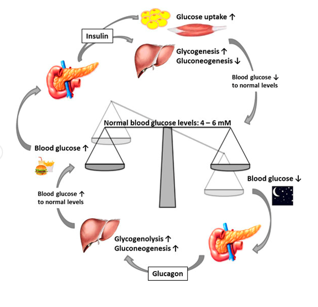
The food's carbohydrate breaks down into simple sugar while you're consuming it. The digestive system may quickly absorb these simple carbohydrates into the blood, which results in a greater blood glucose level (Barclay, Cooper, Ginic-Markovic & Petrovsky, 2010). The pancreas assists in identifying the rising glucose molecules in this circumstance and secretes insulin in response. Insulin aids in controlling and regulating carbs and monitoring the metabolism of fat.
When insulin is released into the bloodstream, it instructs the fat, muscle, and skeletal cells to absorb the blood's glucose molecules. Insulin release from the pancreas ceases when the blood glucose level falls and reaches a safe level.
A person is never able to express or feel how their blood sugar is doing since they cannot sense hyperglycemia. In some circumstances, a person may experience extreme hunger or thirst, urinate more frequently, or even lose consciousness. For diabetics who continue to take insulin, rapid hyperglycemia could result in a hazardous condition (Paine, Pithawalla & Naworal, 2019).
There are times when going without food for a period can cause the blood's level of Glucose to decline. When such a circumstance occurs, the pancreas responds by releasing glucagon, a separate chemical. Glucagon aids in the liver's conversion of glycogen to Glucose, or Glucose that resembles starch, in the blood. As soon as the blood glucose level returns to a safe level, the release of glucagon ceases.
When it comes to controlling the blood glucose level, insulin and glucagon cooperate. Insulin and glucagon act in opposition to maintaining a normal glucose level. Simple or complex signs of hyperglycemia include feeling poorly or being unconscious, brain damage, or even death (Roder, Wu, Liu and Han, 2016). Harm to the kidneys, the heart, the eye or a nerve, as well as some damage to the hands or feet, are additional complications and symptoms. Once hyperglycemia has occurred, these consequences may take some time to manifest. Understanding the regulation of glucose molecules is made easier by the diagram below:
Structure of Glucose
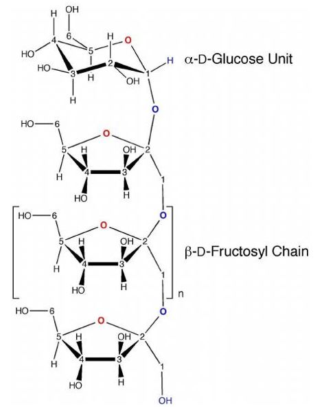
Glucose (C6H1206) is a compound with 6 carbon atoms and an aldehyde group, commonly known as an aldohexose. The presence of glucose molecules might take the shape of an open chain or a ring. The intramolecular interaction between the C atom of the aldehyde and the C-5 hydroxyl group, which results in an intramolecular hemiacetal, creates the ring. When both are present in water, they remain in balance, but when the pH approaches 7, the cyclic one predominates (Paine, Pithawalla & Naworal, 2019). The ring, which has the appearance of a pyran, has 5 carbon atoms and 1 oxygen atom. Cyclic Glucose is also known as glucopyranose. The carbon atoms in the ring are coupled with a side group of hydroxyl to remove the fifth atom, which links back to a carbon atom put outside the ring in the sixth position to produce a group called CH2OH. The illustration below explains the cyclic and acyclic structures of Glucose:
Source: (Barclay, Cooper, Ginic-Markovic and Petrovsky, 2010)
Production of Glucose
Both commercial and natural production of Glucose is possible. The natural process could appear as a byproduct of photosynthesis, which occurs in some prokaryotes and plants. Glycogenolysis is the process by which glycogen breaks down to produce Glucose in both animals and fungi (Barclay, Cooper, Ginic-Markovic and Petrovsky, 2010). In plants, the breakdown manifests as starch. In the liver and kidneys of animals, Glucose is produced.
The commercial procedure can involve the enzymatic hydrolysis of starch to produce Glucose. Certain crops, including maize, potatoes, rice, etc., can be excellent sources of starch. For example, cornflour is frequently utilized in the USA to create glucose molecules from these crops. The enzymatic process occurs in two steps. The enzymes begin to hydrolyze the starch into tiny carbohydrates, which contain glucose molecules in units of five to ten, in one or two hours at 100. C. The starch mixture may occasionally be heated during the process to 130° C or higher (Zhang & Bar-Peled, 2019). The water is heated to assist in dissolving the starch, but heating also deactivates the enzymes, necessitating the addition of additional fresh enzymes after each heating.
The second stage, known as saccharification, uses the glucoamylase enzyme, which is derived from the fungus Aspergillus niger, to completely hydrolyze the partially digested Glucose into glucose molecules. Ph4.0-4.5, at 60°C, and a concentration of carbohydrates weighing 30–35% are necessary for the reaction. If this situation persists for fourteen days, the starch will convert to Glucose with a 96% yield (Rensburg & Ende, 2018). This method can also convert more glucose molecules, but it will use a more diluted solution, which might not be practical. Through the use of filters, the glucose solution produced by this procedure is cleaned before being stored in an evaporator. After multiple crystallizations, a solid form of Glucose is produced.
Functions of Glucose
The metabolism and biosphere both benefit from the widespread usage of Glucose. Compared to some other hexose sugars, Glucose has a weaker ability to react with some proteins that include an amino group. The process is known as glycation results in the destruction or reduction of the function of several enzymes (Dienel, 2018). Proteins produced through glycation are likely to be the source of many acute diabetes-related problems, including blindness and renal failure, but glucose protein added through enzyme regulation may serve a crucial purpose.
Source of Energy
Whether they are bacteria or people, glucose molecules are a fantastic source of energy for practically all living things. Some cells in the body completely rely on
Glucose to produce energy. Both aerobic and anaerobic respiration can utilize the Glucose. A significant portion of the energy used by humans during aerobic respiration comes from carbohydrates, which offer food energy of at least four kilocalories per gramme. Glycolysis converts Glucose into CO2 and water when it comes into touch with the citric acid cycle (Dienel, 2018). Adenosine triphosphate, a type of energy, is produced (ATP). The blood glucose level is controlled by insulin with the aid of additional processes.
Glucose Contained in the Glycolysis
To generate energy for either aerobic or anaerobic respiration, cells use Glucose. The process begins in the early stages of glycolysis, and the first step in it is the phosphorylation of Glucose with the aid of the hexokinase enzyme, which will subsequently break down and release energy. In order to prevent diffusion outside the cell, Glucose is immediately phosphorylated with the aid of the hexokinase enzyme.
As a helper
Glucose serves as a cofactor in the synthesis of proteins and the metabolism of lipids. Some plants and animals can produce more vitamin C thanks to it. The process of glycolysis aids in modifying Glucose for subsequent use. Additionally, glucose molecules aid in the creation of several compounds, including starch, cellulose, glycogen, etc. One of the types of Glucose is lactose, which is found in milk (Dienel, 2018).
As a Source of Absorption
Glucose can be found in dietary carbohydrates as building blocks, in starch, in glycogen, or in conjunction with another monosaccharide as a source of absorption. Additionally, Glucose directly fuels erythrocytes and brain cells (Rensburg & Ende, 2018). Some of them end up in the muscles and liver, where they are stored as glycogen. Additionally, it enters the fat cells, where it is stored as fat. When the body needs energy, it can be drawn from glycogen, which is then converted back into Glucose.
Quick facts about Glucose
From the French word glucose, which means sweet, came to the name glucose. The prefix ose in the word "glucose molecule" designates a carbohydrate.
It is classified as a hexose since it has six carbon atoms. It comes in both linear and cyclic forms.
It is necessary for red blood cells, muscle cells, and the energy delivery of the human brain. Additionally, it can dissolve in water.
The abundant monosaccharide found all around us serves as an energy source for various earthly creatures. It is present in plants in the form of sugar, which is created during photosynthesis.
Additionally, Glucose can create isomers that are similar chemically but have distinct conformations. While L-glucose can be handled synthetically, D-glucose is processed naturally.
The glucose molecule has the chemical formula C6H12O6, which can alternatively be written as CH2O in its simplest form.
References
Barclay, T.G., Cooper, P.D., Ginic-Markovic , M & Petrovsky, N. (2010) Inulin - A versatile polysaccharide with multiple pharmaceutical and food chemical uses. Journal of Excipients and Food Chemicals, 1(3).
Dienel, G.A. (2018) Brain Glucose Metabolism: Integration of Energetics with Function. Physiol Rev, 99(1).
Paine, J.B., Pithawalla, Y.B & Naworal, J.D. (2019) Carbohydrate pyrolysis mechanisms from isotopic labeling. Part 5. The pyrolysis of D-glucose: The origin of the light gases from the D-glucose molecule. Journal of Analytical and Applied Pyrolysis, 138.
Rensburg, H.C.J & Ende, W.V. (2018) UDP-Glucose: A Potential Signaling Molecule in Plants? Front. Plant Sci.
Roder, P.V., Wu, B., Liu, Y & Han, W. (2016) Pancreatic regulation of glucose molecule homeostasis. Experimental & Molecular Medicine, 48, e219.
Zhang, J & Bar-Peled, L. (2019) How Sweet It Is: Small-Molecule Inhibitors of mTORC1 Glucose Sensing. Cell Chemical Biology, 26(9).
Elisa Test, Its Development, Uses, Procedure and Types Assignment Sample
Question
Task: What is understood by Elisa Test? How did it develop? Procedure to conduct Elisa, its uses and types.
Answer
Introduction
Elisa, an enzyme-linked immunosorbent assay, is regarded as a potent method for isolating and measuring a specific protein from a particular complicated mixture. It is a commonly used technique to determine and find proteins in a specific sample. According to Biology Assignment Help specialists the technique is called an immunoassay because antibodies aid in the detection of the proteins. The Elisa Test is employed as a diagnostic tool in plant pathology and pharmaceuticals. In several sectors, quality control is one of its applications. Indirect, direct, sandwich, and competitive/inhibitory Elisa tests are all variations of the Elisa test. It is regarded as a fundamental, adaptable, quantitative, and sensitive test that aids in determining the concentrations of serum antibody (Vencia, Migone & Vito, 2016). The paper will aid in comprehending the Elisa Test concept, its history, and a discussion of its various forms.
What is Understood by Elisa Test?
The Elisa Test is a device that aids in identifying and quantifying the antibodies that are present in blood. When someone is ill or has a condition, the test is useful for identifying the existence of antibodies in their body. The protein that the body produces in response to dangerous things like antigens is what is known as an antibody. The Elisa Test may occasionally be used as a screening tool before doing any other tests. Engvall and Perlmann first used the phrase in 1971, describing it as a method that helps identify antibodies in a protein sample that has been immobilised in microplate wells (Gandikota, Gandhi & Maisam, 2020). The test aids in measuring glycoproteins and aids in the diagnosis of HIV infection, pregnancy tests, the diagnosis of the chicken pox, zika, and rota viruses, among other things.
Development of Elisa Test
Radioimmunoassy was employed on radioactively labelled antigens and antibodies prior to the creation of the Elisa Test. The presence of any antigen or antibody was employed to detect radioactivity. However, some studies were able to predict some health concerns associated with the use of radioactivity, which prompted researchers to look for alternatives. Two different teams led by Stratis Avrameas and G.B. Pierce created the method known as "enzyme linkage" in 1960. In the same year, Wide and Jerker Porath also published an immunosorbent method. Elisa Test was created as a result of independent studies published by Anton Schuurs and B. van Weemen in the Netherlands and by Peter Perlman and Eva Engvall at the University of Stockholm in Sweden (Gandikota, Gandhi & Maisam, 2020). In the classic Elisa, chromogenic reporters were used together with certain substrates to assist change the colour and signal the presence of a particular antigen or analyte. The new method produced signals using fluorogenic, electrochemiluminescent, and quantitative PCR reporters. When detecting many analytes in a single or cluster of assays and requiring higher sensitivities, the use of advanced reporters is advantageous. The majority of the time, the more recent assays use reporters other than enzymes without altering the basic assay principles, which caused the assays to be classified as Elisas.
Procedure to test Elisa
Testing for Elisa involves no complexities; it is a straightforward process. Before the test is performed, a consent form must be signed, and the doctor will assist in outlining the justification for the test's use. In order to take blood samples, a healthcare professional will scrub the arm with an antiseptic. After that, a band will be placed around the arm to apply pressure to the veins and collect the blood in one location (Hoffstetter, Giffin & Brown, 2018). With the use of a needle that is inserted into the vein, a blood sample will be drawn when the veins inflate with blood. Once the needed volume of blood has been drawn, the blood flow will be stopped by replacing the needle with a tiny bandage. The medical professional will instruct you to keep pressure on the area where the needle was inserted; doing so will help to reduce blood flow. Less discomfort is felt during the sample collection process, however the arm may throb.
A laboratory will then conduct an analysis on the acquired sample. A petri plate that already has the specific antigen of the illness or condition for which the sample was taken will be filled with the blood sample by a medical professional or lab worker.
.png)
Source: (Hoffstetter, Giffin & Brown, 2018)
The sample being placed in the plate for the Elisa test is shown in the image above. Both will unite if the blood already contains antibodies to combat the antigen. In order to check and monitor the interaction between the blood and the antigen, the laboratory worker will add an enzyme to the dish. A change in hue indicates the presence of the disease or condition for which the test was performed. The degree of colour change brought on by the addition of enzyme aids the medical staff in quantifying the level of antibody present.
Uses of Elisa Test and Risks Involved
The test is mostly used to find proteins in the body, though it can also check for antigens. The Elisa Test can assist in identifying hormones, bacteria, viruses, allergens, viral fever, and antibodies that the body produces to fight infections. Additionally, it can aid in locating any agent that tries to infect a person.
Despite the fact that the test is straightforward, the subject occasionally runs the risk of contracting an infection, feeling sleepy, having their blood flow continue, etc. In such circumstances, the doctor must be consulted and kept informed of the situation. The test aids in the identification of Covid 19. If such occurrences arise in the near future, it is also vital to inform the doctor (Kamarehei, Khabiri & Saidijam, 2018).
Result Analysis of Elisa
Depending on the analysis done by the facility conducting the test, the results of the Elisa test may differ. Another element that affects the outcome is the condition or illness. When the report is ready, the doctor will go through the findings and assist in interpreting what they mean. Testing positive occasionally means that the disease or condition doesn't actually exist. False positives and false negatives are possible; the former indicate the existence of a condition when none actually exists, and the latter the non-existence of a condition when it actually does (Kamarehei, Khabiri & Saidijam, 2018). Due to this uncertainty, the elisa test may be repeated on a patient within a few weeks, or the doctor may request that some additional delicate tests be performed in order to confirm the diagnosis.
Types of Elisa Test
Immobilizing the sample antigen in the petri dish is the first step in the Elisa test. Direct absorption on the dish's surface or the assistance of an antibody deposited on the plate can both be used to immobilise the antigen. The test is divided into four categories: competitive, sandwich, indirect, and direct.
.png)
Source: (Lauridsen , Holmetoft & Petersen, 2016)
The illustration above clarifies how various elisa tests operate. The alterations included in the process are used to split the categories. The sandwich elisa test has a higher level of sensitivity and durability, making it a potent elisa assay.
Direct Elisa: Compared to other tests, the procedure of detecting the presence of antibodies is quicker since it involves fewer steps. In this method, the antigen is immediately applied to the microtitre plate wells, and then the enzyme that is designated as the primary antibody that recognises the complimentary antigens is added. The test is less likely to be inaccurate because there are fewer procedures and reagents needed to complete it. Although the method does not call for testing a second antibody, there are some specificity-related drawbacks as well. When compared to other elisa tests, antigen immobilisation has a lower specificity, which results in more background noise (Lauridsen , Holmetoft & Petersen, 2016). It occurs because sample proteins and the target protein on the microtitre plate do not specifically interact. Since all of the target proteins are linked together by enzyme-labeled antibodies, the direct elisa is less versatile. The labor-intensive and time-consuming procedure of labelling primary antibodies can have an impact on the immunoreaction. Since there is no secondary antibody, there is less signal amplification, which lowers assay sensitivity. One may say that this method is employed to examine how the immune system reacts to a particular antigen.
Indirect Elisa Test: A subordinate Elisa test Due to the use of an enzyme-labeled secondary antibody that interacts with the primary antibody, this approach exhibits great sensitivity. Since it uses fewer labelled antibodies than direct elisa, it is seen as being more cost-effective. Because the secondary antibody that has been enzyme-labeled bonds to the other primary antibodies, the indirect elisa is more adaptable. Secondary antibodies with anti-species reactivity are typically polyclonal in origin (Lauridsen , Holmetoft & Petersen, 2016). The cross reactivity between a secondary antibody and a bound antigen, which may produce a greater background noise, is another restriction of the indirect elisa. When the secondary antibody is to be incubated, there is an additional step that must be conducted as part of the test. The procedure takes extra time. The indirect elisa technique aids in calculating the overall amount of concentrated antibody present in a particular sample.
Sandwich Elisa: For this technique, capture and detection antibodies are used in pairs. Either a monoclonal or polyclonal antibody may be used. Every antibody has a high degree of epitope specificity, and it has been discovered that this assay works best with antigens that have two epitopes. The antibody pairs must have matched specificities in order for them to attach to various epitopes and produce reliable results. Elisa is detected using direct and indirect methods as a result of the captured antibody mixing with an antigen. Sandwich ELISA testing is used because antigen quantification occurs in both the upper and lower layers of antibodies (Pereira, Cunha & Fernandes, 2020). Because a sandwich elisa has the tendency to produce accurate but unreliable results, it needs to be verified more frequently. Due to the need for matching pairs of antibodies, the test occasionally takes a long time. Elisa plate must be coated with a captured antibody as the first stage in the sandwich assay process. The addition of a sample antigen to the plate in the second stage is followed by the detection of an antibody. Depending on whether the antibody is enzyme-labeled or enzyme-unlabeled, it will either be a direct sandwich elisa or an indirect sandwich elisa. The secondary enzyme-labeled antibody used in the indirect sandwich elisa is found and introduced to bind the primary unlabeled antibody found. In comparison to direct and indirect elisa techniques, sandwich elisa is a more sensitive method (Pereira, Cunha & Fernandes, 2020). The methodology employs both direct and indirect methods, making it more adaptable when used for detection. The test aids in the analysis of complicated samples that are extremely sensitive and specific because it does not require the pre-purification of antigen. However, there are certain drawbacks to this method that must be taken into account. For example, the elisa kit must be verified in advance for reactivity and detection, which can take time.
Competitive/ Inhibition Elisa: This test is also known as blocking elisa and it utilises a plate/surface assay. Although all other elisa techniques can be adapted to fulfil the standards of competitive elisa, it is known as one of the most difficult assays to do. This technique, which is based on a signal produced by the ensuing interference, aids in estimating the concentration of antibodies or antigens in a specified sample (Sahli, Mouelhi & Tlig, 2018). It demonstrates how a given antigen or antibody competes with a labelled antigen or antibody that has a low concentration. The output signal is inversely correlated to the concentration of antigen in a given sample, with weaker output signalling occurring at greater antigen concentrations. The experiment shows how an antigen coats a microtitre plate. Once the ideal blocking and washing procedure has been accomplished, samples of unknown antigens are added. By including labelled detection antibody and substrates like 3,3',5,5'-Tetramethylbenzidine or TMB, it is dragged. The competitive interaction between the sample and the antigen that binds to the multiwall plates with the primary antibody is one of the crucial processes in this test (Sahli, Mouelhi & Tlig, 2018). When the antigen concentration is high, the output signal will be weak, and when the antigen concentration is low, the output signal will be strong. When an antibody is readily available that is specific to the sample antigen, that is when it should be used. As opposed to the sandwich technique, it aids in the detection of all antigen types, no matter how large or little. Before starting the reaction, the sample must be pre-incubated with another component.
Conclusion
It may be said that the Elisa test aids in the discovery of an antigen or an antibody in a particular sample. It assists in determining whether a person has a condition or not, and if so, whether or not he has an antibody to treat the ailment. There are Elisa test kits on the market that include a plate that has already been coated, a detecting antibody, and other chemicals needed to conduct the test. Sandwich elisa tests are among the several types of Elisa tests, and they are thought to be a suitable technique.
References
.png)

- Assignment - Child Care
- Assignment - Mathematics
- Assignment - Accounting
- Assignment - Auditing
- Assignment - Biology
- Assignment - Law
- Assignment - Management
- Assignment - Nursing
- Assignment - Finance
- Assignment - Computer Science and IT
- Assignment - Humanities
- Assignment - Economics
- Assignment - Statistics
- Assignment - Architecture
- Assignment - Engineering
- Assignment - cookery
- Assignment - Marketing
- Case Study - Chemistry
- Case Study - Accounting
- Case Study - Law
- Case Study - Management
- Case Study - Nursing
- Case Study - Finance
- Case Study - Computer Science and IT
- Case Study - Engineering
- Case Study - Economics
- Case Study - Biology
- Case Study - Auditing
- Case Study - Marketing
- Case Study - Project Management
- Coursework - Diploma
- Coursework - Accounting
- Coursework - Auditing
- Coursework - Biology
- Coursework - Management
- Coursework - Nursing
- Coursework - Finance
- Coursework - Computer Science and IT
- Coursework - Engineering
- Coursework - Humanities
- Coursework - Child Care
- Coursework - Project Management
- Coursework - Economics
- Coursework - Cookery
- Coursework - Law
- Dissertation - Accounting
- Dissertation - Auditing
- Dissertation - Biology
- Dissertation - Law
- Dissertation - Management
- Dissertation - Nursing
- Dissertation - Finance
- Dissertation - Computer Science and IT
- Dissertation - Humanities
- Dissertation - Economics
- Essay - Politics
- Essay - Childcare
- Essay - Accounting
- Essay - Biology
- Essay - Law
- Essay - Management
- Essay - Nursing
- Essay - Computer Science and IT
- Essay - Humanities
- Essay - Economics
- Essay - Auditing
- Essay - Engineering
- Essay - Architecture
- Essay - Finance
- Essay - Science
- Essay - Marketing
- Programming - Computer Science and IT
- Reports - Management
- Reports - Computer Science and IT
- Reports - Project Management
- Reports - Marketing
- Reports - Nursing
- Reports - Engineering
- Reports - Accounting
- Reports - Humanities
- Reports - Finance
- Reports - Architecture
- Reports - Biology
- Reports - Economics
- Reports - Childcare
- Reports - Law
- Research - Accounting
- Research - Auditing
- Research - Biology
- Research - Law
- Research - Management
- Research - Nursing
- Research - Finance
- Research - Computer Science and IT
- Research - Science
- Research - Engineering
- Research - Humanities
- Research - Economics
- Research - Project Management
- Research - Statistics
- Research - Architecture
- Research - Marketing
- Thesis Writing - Computer Science and IT
- Thesis Writing - Engineering
- Thesis Writing - Biology
- Thesis Writing - Finance
- Thesis Writing - Humanities
- Thesis Writing - Auditing
- Thesis Writing - Economics
- Thesis Writing - Law
- Thesis Writing - Nursing
- Thesis Writing - Accounting
- Thesis Writing - Architecture


.png)
~5.png)
.png)
~1.png)


.png)